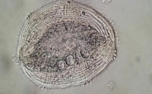
Kentrophyllum verrucosum.
Kentrophyllum verrucosum from the shoreline benthos of Lion Head marina at the mouth of marine estuary Hog Creek where it empties into Gardiner's Bay. This species is sufficiently distinctive with its marginal trichocystic warts around the entire circumference of the body margin each associated with three or four "soft" spines to identify it as Kentrophyllum verrucosum. Thus far I have found two individuals measuring 200 um and 150 um. They tend to expand and contract becoming ellipsoid to more rounded in shape.
Imaged in Nomarski DIC using Olympus BH2 under SPlan 40x and 20x objectives plus phone cropping on Samsung Galaxy S9+.
Members of the order Pleurostomatida are haptorians with a distinctive, laterally compressed, flattened body that are common in a variety of cosmopolitan biotopes. There are over 200 morphospecies assigned to 12 genera within two families reported from a variety of aquatic environments in Europe, Asia, North America, and even Antarctica. In the pleurostomatid family Amphileptidae, the relatively small genera Kentrophyllum and Epiphyllum comprise fewer than 10 morphospecies differing from other amphileptids by having a large number of somatic kineties forming sutures on both right and left sides and an extremely elongate perioral kinety at the periphery of the body. A single morphological character (absence of spines surrounding the margin of the cell) distinguishes Epiphyllum from Kentrophyllum.
The classification history is as follows:
Kentrophyllum verrucosum (Stokes, 1893) Petz et al., 1995. Synonymy.
Litosolenus verrucosus in Stokes, 1893, p. 302 (original description), Loxophyllum setigerum in Sauerbrey, 1928, p. 369 (misidentification), Loxophyllum verrucosum in Kahl, 1931, p. 201 (new combination), Loxophyllum verrucosum in Kahl, 1933, p. 63 (description of German population), Kentrophyllum verrucosum in Petz et al., 1995, p. 55 (new combination).
Description of population from Zhanjiang. Cells highly variable in size, measuring 125–250 μm × 65–185 μm in vivo, usually 170–200 μm × 90–150 μm. Body ranging in shape from slender when swimming to nearly oval when stationary (Fig 4B–4D and 4G). Length to width ratio 3–4:1. Margin of cell flat, thin, hyaline, ~10 μm wide (Fig 4B). Three to 7 (usually 4) macronuclear nodules located near center of cell; nodules spherical to ellipsoid, measuring 10– 15 μm × 10–12 μm in vivo (Fig 4C, 4D and 4G). Micronucleus not observed. Four to 7 contractile vacuoles, with diameter of 5–12 μm, located in dorsal half of cell (Fig 4C, 4D and 4G). Extrusomes bar-shaped, ~15 μm long, always clustered to form 6–19 warts along margin of body excluding oral area; interval between warts usually concave. Each wart with 1–4 (usually 3) soft spines, each spine thick at base and tapering to sharp point; one spine in each cluster always much longer (6–7.5 μm) than others (3–4.5 μm) (Fig 4A, 4C, 4E, 4G and 4K). Pellicle thin; cytoplasm gray to dark in center of cell, depending on number of food vacuoules present, and often containing numerous, refringent globules measuring 2–5 μm in diameter. Right side flat with many inconspicuous, longitudinal, shallow grooves (Fig 4G); left side slightly to distinctly arched, with many conspicuous, longitudinal, shallow grooves (Fig 4F). Thirty to 50 peribuccal papillae densely arranged along left edge of oral slit (Fig 4H). Movement by left side of body slow gliding on substrate, usually stationary, rarely swimming in water column.
Right-hand somatic kineties densely ciliated, numbering 38–45 in (including PK2) and forming a distinct anterior suture and an inconspicuous posterior suture. Left side of body also densely ciliated, with 28–33 somatic kineties (including PK1 and dorsal brush kinety) forming a distinct suture at both anterior and posterior ends of the body. Dorsal brush kinety extending half of body length, formed of densely spaced dikinetids. Nematodesmata well-developed, up to 150 μm long.
Discussion and supporting figures adapted from:
Phylogenetic and Taxonomic Revision of an Enigmatic Group of Haptorian Ciliates, with Establishment of the Kentrophyllidae fam. n. (Protozoa, Ciliophora, Litostomatea, Pleurostomatida). Lei Wu, John C. Clamp, Zhenzhen Yi, Jiqiu Li, Xiaofeng Lin. DOI:10.1371/journal.pone.0123720 May 6, 2015