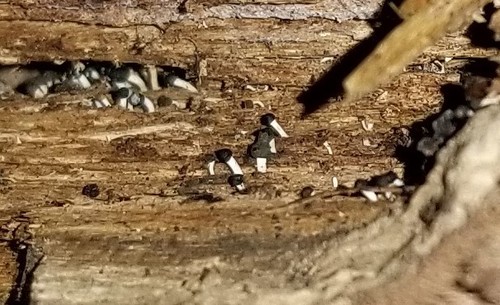
Growing on the hollowed-out inside of a corticate Acer sacchariferum stick. The “heads” of some of the synnemata were being eaten by millipedes. Clamped hyphae in the base of the synnemata with lateral “nubs” that may have been the starts of blastoconidia or “secretory cells”. Brown conidia breaking off from clamped hyphae (arthroconidia) in the “heads” of the synnamata within a drop of slime. Conidia measurements from Piximetre: (11.6) 12.5 – 19 (23.9) × (4.8) 5.2 – 6.5 (7.1) µm, Q = (1.9) 2 – 3.5 (5); N = 30, Me = 16.2 × 5.8 µm; Qe = 2.8
Individual conidia: 16.94 × 5.91 µm, 12.83 × 6.48 µm, 16.69 × 6.55 µm, 13.60 × 5.93 µm, 14.11 × 5.80 µm, 15.73 × 6.06 µm, 11.90 × 5.12 µm, 12.98 × 6.82 µm, 15.12 × 7.15 µm, 16.32 × 5.72 µm, 13.73 × 5.61 µm, 11.58 × 6.09 µm, 11.95 × 5.84 µm, 14.72 × 5.07 µm, 19.02 × 6.23 µm, 18.22 × 5.47 µm, 17.87 × 5.17 µm, 17.45 × 6.34 µm, 18.37 × 5.42 µm, 19.05 × 5.26 µm, 15.82 × 6.27 µm, 17.72 × 5.71 µm, 16.88 × 5.67 µm, 16.51 × 5.35 µm, 17.55 × 5.49 µm, 17.44 × 5.35 µm, 23.85 × 4.78 µm, 18.33 × 5.91 µm, 12.52 × 5.35 µm, 22.65 × 5.80 µm