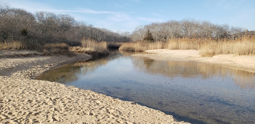
Chaenea mirabilis Kwon et al., 2014. The ciliates appeared in a bacterial biofilm that formed after three days associated with decaying Ectocarpus species that had formed small floating tufts in the saline channel (23.4% salinity) between marine bay Napeague Harbor and Fresh Pond. After two days they had disappeared and on day two individuals were fewer and smaller. Individuals ranged from 100 up to 125 um in length.
"The widespread haptorid genus Chaenea Quennerstedt, 1867 has been found in marine sand, freshwater, brackish water and moist soil. Its members are characterized by the following features: cell elongate and contractile; cytostome apically located and surrounded by dikinetid circumoral kinety; somatic kineties which are slightly spiralled when contracted and mainly composed of monokinetids; dorsal brush consisting of four dikinetidal rows; one permanent contractile vacuole located at the posterior end of the body; extrusomes rod-like or thorn-like, attached to the oral bulge and scattered in the cell. Since being established, 14 nominal species have been assigned to this genus, namely, C. crassa Maskell, 1887, C. gigas Kahl, 1933, C. limicola Lauterborn, 1901, C. minor Kahl, 1926, C. mirabilis Kwon et al., 2014, C. psammophila Dragesco, 1960, C. robusta Kahl, 1930, C. sapropelica Kahl, 1930, C. simulans Kahl, 1930, C. stricta (Dujardin, 1841) Foissner et al., 1995, C. teres (Dujardin, 1841) Kent, 1881, C. tesselata (Kahl, 1935) Dragesco and Dragesco-Kerneis, 1986, C. torrenticola Foissner, 1984, and C. vorax Quennerstedt, 1867. In 1995, Foissner et al. synonymized C. torrenticola with C. stricta. Consequently, 13 species and an unidentified species from Petz et al. (1995) remain in the genus. Among these species, most have only been reported once, mainly based on living observation. Data on the ciliary pattern, especially detailed information regarding the dorsal brush, is only available for C. teres and C. mirabilis (Kwon et al. 2014, Petz et al. 1995)" (2).
Chaenea mirabilis was described by Kwon et al in 2014, from brackish water collected near the town of Busan, Korea. "Chaenea mirabilis is distinguished from all congeners by the combination of the following traits: (i) a narrowly bursiform to flask-shaped, 60–100 um long body; (ii) 11–21 doughnut-shaped or sometimes horseshoe-shaped macronuclear nodules; (iii) two types of extrusomes: type I is rod-shaped and 6–8 um long, while type II is narrowly to broadly teardrop-shaped and only 1.5–2 um long; (iv) highly refractive special granules tightly arranged between the first and second brush row, forming a conspicuous bulge; and (v) 12–13 somatic kineties" (1).
"Among congeners, C. mirabilis sp. n. is outstanding in having highly unique refractive granules that form a conspicuous bulge between brush rows 1 and 2. Another peculiar feature of this species is the doughnut-shaped or sometimes horseshoe-shaped macronuclear nodules. These have, however, also been found in one other species, C.
minor, by Kahl (1926). Both species are similar in body size (50–60 um in C. minor and 60–100 um in C. mirabilis), but can be distinguished by body length:width ratio (3:1
vs. 7:1), number of macronuclear nodules (5–6 vs. 11–21), and habitat (shallow puddle filled with leaf-litter vs. brackish water). The 18S rRNA gene sequence of C. mirabilis is very similar to that of C. teres and C. vorax. However, the two latter species differ distinctly from C. mirabilis in that they have a much larger body (100–400 um in
C. teres and 100–180 um in C. vorax vs. 60–100 um in C. mirabilis) and by the shape of the macronuclear nodules (globular to narrowly ellipsoidal in C. teres and C. vorax vs. doughnut- or horseshoe-shaped in C. mirabilis). Moreover, the conspicuous, highly refractive subcortical globules of C. mirabilis have not been observed in C. teres
or C. vorax (Foissner 1984; Kahl 1930; Song and Packroff 1997; Song et al. 2009).
My observation is somewhat larger than the size range of the Korean population measuring 100-125 um in length. However, the nodular collection of such granules between the first and second brush row is usually very species-specific (Peter Vd’acny-personal communication) and the finding of horseshoe and donut shaped macronuclear nodules makes the diagnosis of Chaenea mirabilis Kwon et al., 2014 quite secure. I believe I have also demonstrated the two types of extrusomes E1 and E2.
I encountered an interesting vignette of life in the protist jungle involving Chaenea mirabilis, a tug of war between a Chaenea who had stolen and ingested a small protist that had been prey to a suctorian who was hanging on by its tentacle to its stolen meal until the Chaenea snapped the suctorian's grip.
-
Morphology and Molecular Phylogeny of a New Haptorian Ciliate, Chaenea mirabilis sp. n., with Implications for the Evolution of the Dorsal Brush in Haptorians (Ciliophora, Litostomatea) Choon Bong Kwona , Peter Vd’acny , Shahed Uddin Ahmed Shaziba & Mann Kyoon Shin. Journal of Eukaryotic Microbiology 2014, 61, 278–292
-
Morphology of Two Novel Species of Chaenea (Ciliophora, Litostomatea): Chaenea paucistriata spec. nov. and C. sinica spec. nov. Xinpeng FAN, Yuan, Fukang GU, Jiqiu LI, Saleh A. AL-FARRAJ, Khaled A. S. AL-RASHEID and Xiaozhong HU. Acta Protozool. (2015) 54: 97–106

